


Pediatric neurology focuses on diagnosing and managing brain, spinal cord, nerve, and muscle disorders in infants, children, and teens. The scope includes epilepsy, developmental delays, headache, movement issues, neuromuscular conditions, and inflammatory disorders. As tools improve, patterns become easier to detect, and findings get linked to a child’s stage of growth. Here are some recent advancements in this field:
Electroencephalography
Electroencephalography (EEG) records brain waves using small sensors on the scalp, tracking changes that happen in fractions of a second. This imaging tool is especially helpful for children, as it aids in analyzing seizures, sleep stages, and attention-related episodes. Movement can create noise in the readings, but modern filters help reduce this, and technicians coach children to stay relaxed during the test. Clinicians examine background rhythms, symmetry between the two sides of the brain, and spikes that may indicate seizure risk. They also test how the brain responds to light or deep breathing.
Different EEG setups serve different diagnostic needs. In newborns, trend-based bedside monitoring tracks sleep and wakefulness over extended periods of time. Since brain rhythms change with age, reference charts by month and year aid in interpretation. The results help determine seizure type, possible starting points in the brain, and guide follow-up plans.
Magnetic Resonance Imaging
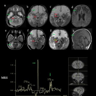
Magnetic resonance imaging (MRI) utilizes magnets and radio waves to produce clear images of the brain and spine, eliminating the need for ionizing radiation exposure in patients. Advanced MRI techniques further enhance these images by mapping the movement of water through tissues. When high-resolution photos are combined with these specialized maps, healthcare providers can detect subtle differences that correspond to symptoms or learning challenges. Strategies in pediatric neurology may enhance scan quality, including child-friendly coaching, shorter protocol times, and motion control techniques to minimize the need for repeat scans.
Computed Tomography
Computed tomography (CT) scans use X-rays to create detailed cross-sectional images of the body, and their speed is one of their main advantages. When a head injury occurs, quick scanning can identify fractures or bleeding. Settings may be adjusted based on the patient’s age to minimize radiation dose. While MRI provides more detailed images of soft tissues, CT scans are particularly effective for viewing bones, calcifications, and fresh blood. Medical teams look at factors such as timing, access to equipment, and whether the child can remain still during the scan. Different clinical situations require different CT techniques. When deciding which imaging tools to use, take the following steps:
- Request a written plan that includes the following: a list of tests, home monitoring tips, and contact information for after-hours assistance.
- Discuss rehabilitation options, such as physical, occupational, or speech therapy, when function or daily tasks change.
Find Pediatric Neurology Treatments
Treatment planning starts with accurate information and steady follow-up. Bring a timeline of symptoms, any videos of events, and a complete list of medicines and supplements. Ask about goals for home tracking, what changes to look out for, and when to follow up. Stay organized with a folder that includes reports, lab results, and imaging summaries. If you’re ready to take the next step, reach out to a pediatric neurology clinic. Share your child’s history, schedule an evaluation, and ask which tests fit your concerns. Call today to schedule an appointment and establish a clear path for assessment and follow-up.
- Braces for Adults and What You Need to Know
- Benefits of Invisible Braces for Teens and Adults
- Understanding the Link Between Behavioral Studies and Mental Health Support Services
- Key Characteristics That Support Long-Term Success in Nursing
- How Continued Nursing Education Supports Safer and More Effective Healthcare


Leave a Reply